Have questions about your insurance coverage?
PET/CT
PET/CT Overview
Advanced PET/CT Imaging for More Complete Answers
PET/CT is an advanced imaging technology that combines two powerful imaging technologies to allow your specialized radiologists to see both what is happening inside your body and exactly where it is happening.
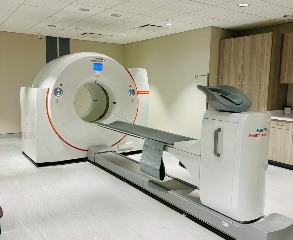
Expert PET/CT care in Northern Virginia
At Fairfax Radiology Centers, our PET/CT examinations are performed by experienced technologists and interpreted by specialty-trained radiologists with expertise in PET/CT and molecular imaging.
We offer a range of PET/CT examinations using specialized radiotracers to meet different clinical needs.
Our goal is to provide advanced imaging, expert interpretation, and compassionate care throughout your imaging experience—while providing your referring physician with the information needed to help guide your care.
Procedures
Beta-Amyloid PET/CT Scan
A Beta-Amyloid PET/CT scan is an advanced, non-invasive brain imaging test used to evaluate memory loss, cognitive decline, and suspected Alzheimer’s disease.
FDG Brain PET/CT (Metabolic Function)
A FDG brain PET/CT is an advanced imaging test that evaluates how the brain uses glucose, providing information about brain function and metabolism.
PET/CT Body Scan
A PET/CT body scan is an advanced, non-invasive imaging test used to detect, evaluate, and monitor disease throughout the body.
PSMA PET/CT with PYLARIFY
A PET/CT PSMA scan is an advanced imaging test used to detect and evaluate prostate cancer throughout the body. It combines two imaging technologies to provide detailed information about the location and activity of prostate cancer cells.

What is a PET/CT scan?
A PET/CT scan combines Positron Emission Tomography (PET) with Computed Tomography (CT) in a single examination.
PET shows how tissues and cells are functioning. A small amount of a radiotracer is given through an intravenous (IV) line. The radiotracer travels through the body and collects in areas based on the specific biological process it is designed to show. The PET scan detects this activity and creates images that can help identify areas of abnormal activity.
CT provides detailed pictures of the body's anatomy.
By combining PET and CT images, your specialized radiologist can see both areas of activity and exactly where they are located, providing a more complete picture than either scan alone.
Why is PET/CT performed?
PET/CT is most commonly used to help detect, locate, stage, and monitor cancer. It can also help evaluate certain neurological and other medical conditions, depending on the type of radiotracer used.
Different radiotracers are designed to show different biological processes. Your physician will select the appropriate PET/CT examination based on your medical condition and the questions your physician needs answered.
What can I expect during a PET/CT?
A small IV will be placed in your arm to give you the radiotracer. You will then have a resting period while the radiotracer travels through your body and reaches the tissues being evaluated.
Subsequently, you will lie on the scanning table while it moves through the PET/CT scanner. The scan is painless and noninvasive. The exact preparation, resting period, and scan time depend on the type of PET/CT examination you are having.
Why Would My Physician Recommend a PET/CT?
PET/CT may be recommended to:
- Help detect and locate disease
- Determine the extent or stage of certain cancers
- Assess how a disease is responding to treatment
- Provide additional information when evaluating certain neurological conditions
- Help guide physicians in diagnosis and treatment decisions
How Does a PET/CT Scan Work?
Before the examination, a small amount of a specialized radiotracer is administered, usually through an intravenous (IV) line. The radiotracer travels through the body and collects in specific tissues or areas of interest.
The PET portion of the examination detects signals from the radiotracer to show biological or metabolic activity, while the CT portion provides detailed anatomical images. Together, the images give radiologists information about both the function and structure of tissues and organs.
Because PET/CT exams are performed for different clinical purposes, the type of radiotracer, preparation, use of contrast, uptake time and examination length can vary. Your imaging team will provide specific instructions based on the type of PET/CT you are having.