Thyroid nodules are more common than many people realize. According to the American Thyroid Association, up to 50% of adults will develop a thyroid nodule during their lifetime. While most of these nodules are benign and do not cause symptoms, it's important to accurately evaluate them to ensure peace of mind and appropriate medical care.
What Is a Thyroid Nodule?
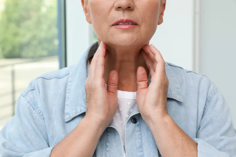
A thyroid nodule is a growth or lump that develops within the thyroid gland, located at the base of the neck. Nodules can be solid or filled with fluid and are often discovered during a physical exam or incidentally through imaging for another concern. Most do not cause noticeable symptoms, though some may lead to swelling or discomfort in the neck, difficulty swallowing, or voice changes.
How Are Thyroid Nodules Evaluated?
At Fairfax Radiology Centers, the evaluation of a thyroid nodule typically begins with a thyroid ultrasound—a safe, non-invasive imaging exam that helps determine the size, shape, and characteristics of the nodule. Based on the ultrasound findings, the radiologist may recommend a Fine Needle Aspiration (FNA) biopsy to collect a small sample of tissue for further analysis. This minimally invasive procedure helps determine whether the nodule is benign or potentially cancerous.
What Should I Expect During a Neck/Thyroid Ultrasound?
If this is your first ultrasound, you may not be sure what to expect. Not to worry, we are here to guide you every step of the way. During a Neck/Thyroid Ultrasound, warm gel will be applied to the neck area to be examined. A probe is gently moved back and forth on the skin over the area, using sound waves to create an image of the thyroid on a screen. This procedure is quick, painless, and non-invasive.
What Should I Expect After a Neck/Thyroid Ultrasound?
Once the ultrasound is complete, the gel will be wiped off your skin. Any gel that may be left behind will dry like powder and will not stain or discolor your clothing. You can resume your normal activities immediately following the procedure.
What Can I Expect During an Image-Guided Thyroid Biopsy?
If the ultrasound shows any suspicious or unclear areas, a Fine Needle Aspiration (FNA) biopsy may be recommended to further evaluate the thyroid nodule.
There is typically no special preparation needed for this procedure. Before the biopsy begins, your neck will be cleansed. An ultrasound transducer is placed on your neck to locate the thyroid nodule. Using ultrasound guidance, a subspecialized radiologist will insert a fine needle through the skin and into the nodule to collect a tissue sample.
It’s important to remain still during the procedure. If multiple nodules need to be tested, the process will be repeated for each one. Once all samples are collected, pressure will be applied to the biopsy area to stop or prevent any bleeding, and a bandage may be placed if needed.
What Can I Expect After the Biopsy?
The collected tissue samples will be sent to pathology for analysis. Your physician will receive a report and discuss the results with you, along with any next steps. You can resume normal activities after the procedure. Mild soreness at the biopsy site is common and usually resolves within a day or two.
Comprehensive Thyroid Imaging at FRC
Fairfax Radiology Centers offers both thyroid ultrasound and FNA biopsy services, performed by a team of subspecialized radiologists with advanced expertise in diagnostic imaging and image-guided procedures. Our goal is to provide patients with accurate information, compassionate care, and confidence throughout the diagnostic process.
While only a small percentage of thyroid nodules are cancerous, early and precise evaluation is key. If you’ve been referred for thyroid nodule evaluation or are experiencing symptoms, FRC is here to support you every step of the way.